Digital Dental Scans: Why We Skip the Goopy Impressions
If your memory of dental impressions involves a tray full of putty, a few long minutes of holding still and trying very hard not to gag, there is some good news: many dental records can now be captured digitally.
A digital dental scan uses a small intraoral scanner to record the visible surfaces of your teeth and surrounding oral structures. As the scanner moves around your mouth, software builds a detailed three-dimensional model that the dental team can view on screen.
At TOOTHbar in southwest Calgary, digital scanning is part of a broader tech-forward approach to treatment planning. Scans can support restorative dentistry, orthodontics and other custom dental work while also giving you something traditional impressions never did very well: an immediate, enlarged view of your own teeth.
Here is how the technology works, what it can be used for, how it compares with conventional impressions, and where scans fit alongside dental X-rays and CBCT imaging.
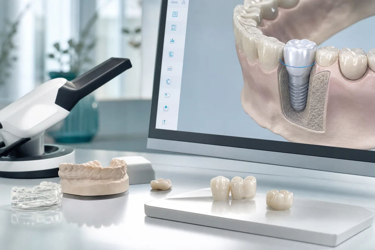
A digital dental scan creates a three-dimensional model that can become part of the workflow for custom restorative and orthodontic treatment.
In this guide
- What is a digital dental scan?
- How does an intraoral scanner work?
- What does the scan feel like?
- Digital scans vs putty impressions
- Are digital scans accurate?
- What are digital dental scans used for?
- Digital scans for orthodontics
- Crowns and restorative dentistry
- Digital scans and implant planning
- Is a 3D dental scan the same as an X-ray?
- Does an intraoral scan use radiation?
- Are traditional impressions completely gone?
- Digital dentistry at TOOTHbar
- Frequently asked questions
What Is a Digital Dental Scan?
A digital dental scan is a digital record of the visible surfaces inside your mouth. Instead of filling an impression tray with material and waiting for it to set, the clinician uses a handheld device called an intraoral scanner.
The scanner captures many images or data points as it moves across the teeth. Software combines that information into a three-dimensional digital model of your dental arches and bite.
The digital model can be rotated, enlarged and viewed from different angles during planning and discussion.
For procedures suitable for digital impressions, there is no need to wait while a full tray of conventional impression material sets.
The scan can be reviewed immediately and incorporated into compatible restorative or orthodontic workflows.
An intraoral surface scan is not a replacement for dental X-rays or CBCT imaging when information beneath the visible surface is required.
How Does an Intraoral Scanner Work?
The scanner is moved systematically around your upper teeth, lower teeth and bite. Optical imaging technology records the surfaces as the wand moves.
The computer then combines the captured information into a virtual dental model.
One useful advantage happens before the scan is even finished
The clinician can inspect the digital model on screen and identify an area that may need additional information.
Instead of necessarily starting an entire impression again, the missing or unclear area can often be rescanned and incorporated into the digital record.
The Canadian Dental Association has previously described digital impression workflows in which the clinician captures the preparation, surrounding teeth, opposing arch and bite before reviewing the three-dimensional image and sending the digital information into the restorative workflow.
For additional background, see the Canadian Dental Association discussion of digital impression workflows .
What Does a Digital Scan Feel Like?
You sit in the dental chair while the clinician guides the scanner around your mouth. You may be asked to move your tongue or turn slightly so different surfaces can be captured.
There is still a device in your mouth, so calling the process completely sensation-free would be unrealistic. The major difference is that there is no large tray filled with impression material sitting in place while the material sets.
That can be particularly welcome for people who have disliked the taste, bulk or gagging sensation associated with traditional impressions.
What does the research say about comfort?
Studies comparing digital and conventional impression techniques generally report higher patient preference and less discomfort with intraoral scanning. A systematic review and meta-analysis found that patients preferred intraoral scans and reported less discomfort, nausea, unpleasant taste and breathing difficulty than with conventional impressions.
See the PubMed-indexed systematic review of patient experiences with digital and conventional impressions .
Digital Dental Scans vs Traditional Putty Impressions
| Difference | Digital Intraoral Scan | Conventional Impression |
|---|---|---|
| How the record is captured | A handheld optical scanner records dental surfaces and creates a digital model. | Impression material is placed in a tray and allowed to set around the teeth. |
| Record produced | Three-dimensional digital file. | Physical negative impression that can be used to create a model. |
| Review | The digital model can generally be inspected on screen during the appointment. | The physical impression is inspected after removal. |
| Missing information | Specific areas can often be rescanned. | A significant defect may require another impression. |
| Patient experience | Generally preferred by patients in comparative studies. | Requires a tray and impression material to remain in the mouth while setting. |
| Clinical use | Useful for many orthodontic and restorative workflows. | Still useful and sometimes preferred or required in particular clinical situations. |
Are Digital Dental Scans More Accurate?
This deserves a more careful answer than simply saying digital is always better.
Modern intraoral scanners can create highly detailed records and are well established for many orthodontic and restorative applications. The clinician also has the advantage of being able to inspect the digital record immediately and rescan areas that need more information.
But accuracy depends on the scanner, the clinical situation, the size of the area being captured, operator technique and the treatment being planned.
Research comparing digital and conventional impressions for tooth-supported restorations has found digital techniques to be clinically comparable in areas such as marginal fit while offering better patient comfort.
A systematic review and meta-analysis published in the Journal of Prosthetic Dentistry found digital and conventional impressions comparable for marginal fit in the tooth-supported restorations studied, while digital scanning scored higher for patient comfort.
Digital does not mean appropriate for every possible case
Certain complex situations can still favour conventional impression techniques. The dentist chooses the record-taking method based on what provides suitable information for the treatment being planned.
What Are Digital Dental Scans Used For?
One reason intraoral scanning has become such a useful dental tool is that the same type of three-dimensional surface record can support several different workflows.
Digital Scans for Invisalign and Orthodontics
Orthodontics is one of the most familiar uses for an intraoral scanner.
A digital model records the current position of the teeth and the way the upper and lower arches relate. That information can become part of diagnosis, treatment planning and a digital orthodontic workflow.
TOOTHbar currently offers Invisalign clear aligners as well as fixed orthodontic options, and digital records form part of its technology-forward approach to treatment planning.
If you are considering tooth movement, learn more about orthodontics at TOOTHbar.
A scan is a record, not the diagnosis
The digital model gives the dentist useful information about visible tooth surfaces and relationships, but orthodontic diagnosis can require an examination, photographs, radiographs and other records depending on the individual case.
Digital Scans for Crowns and Restorative Dentistry
Digital impressions are also widely used in restorative dentistry.
When a treatment is suitable for a digital workflow, the scanner can record the prepared tooth, neighbouring teeth, opposing arch and bite relationship. That file can then become part of designing or manufacturing a custom restoration.
TOOTHbar's live restorative page specifically notes that restorative consultations can include diagnostic imaging and a digital scan.
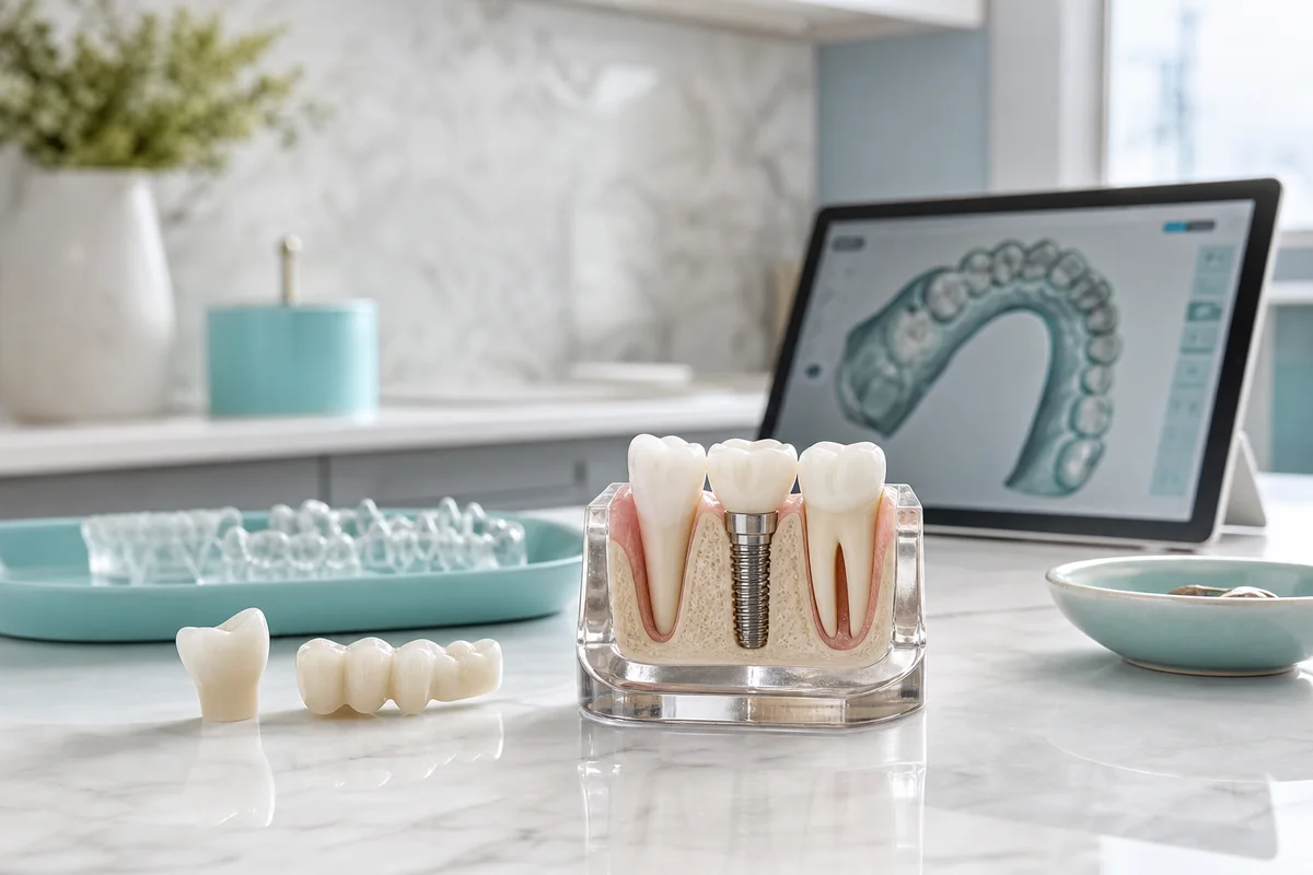
Digital information can move directly into restorative planning
Rather than first turning a physical impression into a stone model, compatible digital workflows allow the scan file itself to become part of the design and laboratory process.
Explore TOOTHbar's restorative dentistry services for fillings, crowns, bridges and other treatment for damaged teeth.
What About Dental Implant Planning?
Implant dentistry is a good example of why the phrase "3D dental scan" can become confusing.
An intraoral scanner captures the visible surfaces of the mouth. Implant planning may also require CBCT imaging, which provides three-dimensional information about structures beneath the surface, including bone.
These are different technologies that can complement one another in a digital workflow.
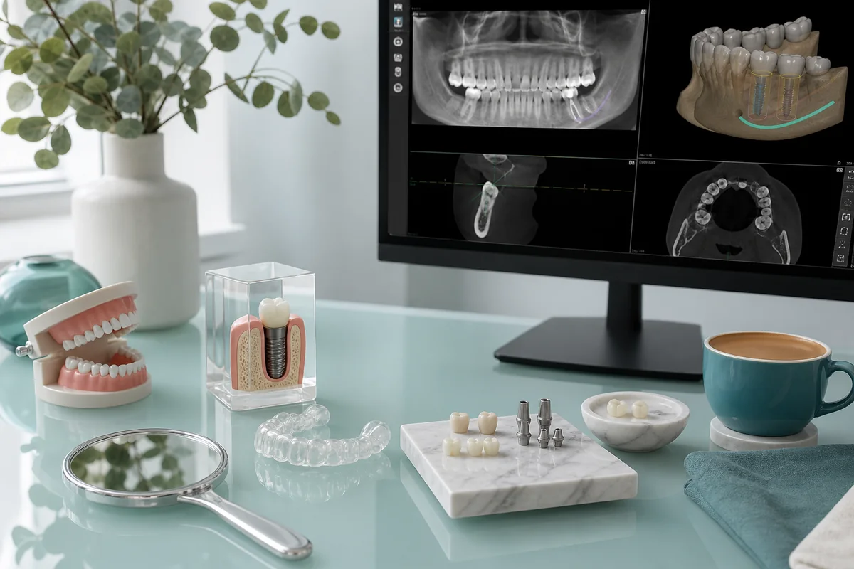
Intraoral scanning and CBCT imaging answer different clinical questions. Implant planning may use information from both.
TOOTHbar performs CBCT evaluation on site for implant and restorative cases when indicated. You can learn more about dental implants at TOOTHbar.
Does a Digital Dental Scan Replace X-Rays?
No. They show different information.
An intraoral surface scan is excellent at creating a three-dimensional model of surfaces the scanner can see. Dental radiographs are used when the dentist needs information that cannot be understood from the visible surface alone.
Records the visible shape and position of teeth and other scan-accessible oral surfaces.
Can provide information about structures that cannot be evaluated simply by looking at the visible surface.
Can provide volumetric information about teeth, bone and surrounding structures when clinically indicated.
Your dentist selects imaging and records based on what is necessary for diagnosis and treatment planning.
Does a Digital Dental Scan Use Radiation?
The standard optical surface-scanning function of an intraoral scanner does not use dental X-ray radiation to create the three-dimensional surface model.
That does not mean a scan eliminates the need for radiographs. If your dentist needs X-ray information for diagnosis or treatment planning, those images serve a different purpose.
Some modern intraoral scanning systems can also incorporate additional optical technologies, but those features vary by scanner and should not be confused with conventional dental radiographs or CBCT.
Current intraoral scanner information from iTero explains how optical scanning captures detailed three-dimensional dental impressions and describes additional imaging features available on certain scanner configurations.
Are Goopy Dental Impressions Completely Gone?
Not completely.
Digital scanning has replaced conventional impressions for many applications, but dentistry is not a situation where one tool is automatically appropriate for every mouth and every procedure.
Research has identified situations where conventional techniques may still be useful or more appropriate. For example, accurately recording large areas of movable soft tissue or certain fully edentulous arches can present additional challenges for intraoral scanners.
The goal is not to use technology for technology's sake
The right record is the one that gives the dental team the information required for your treatment. Most patients may encounter digital scanning far more often than they did in the past, but there are still legitimate reasons a dentist might occasionally choose a conventional impression.
One of the Biggest Benefits Is Being Able to See Your Own Teeth
Digital scanning is not only about sending information to a lab.
When your dental model appears on screen, you can look at the same general record your dentist is discussing. Crowding, tooth position, wear, spacing and the shape of existing restorations can become much easier to understand when you can rotate and enlarge the model.
That fits especially well with TOOTHbar's focus on co-creating treatment plans rather than asking patients to simply accept a recommendation they do not understand.
Better technology should make the conversation clearer
A sophisticated scan is only useful if it helps the clinical team plan care or helps you understand what is being discussed. The screen should support the conversation, not replace it.
How Digital Scans Fit Into Cosmetic Dentistry
Cosmetic treatment often depends on understanding proportions, tooth shape, bite relationships and the space available for any planned changes.
Digital models can contribute to planning for treatments such as veneers, restorative work and selected smile-design workflows. Depending on the treatment and software being used, digital records may also help demonstrate possible changes before treatment begins.
That does not mean a computer preview guarantees an exact final result. Simulations and digital designs are planning and communication tools.
If you are considering changes to your smile, explore cosmetic dentistry at TOOTHbar.
Digital Dentistry at TOOTHbar
Digital scanning fits naturally with the way TOOTHbar approaches dentistry: modern technology should make treatment planning clearer and the patient experience easier without replacing clinical judgement.
The live TOOTHbar restorative process specifically includes diagnostic imaging and digital scanning where appropriate, and digital workflows also support the studio's orthodontic, implant and cosmetic services.
For patients, that can mean fewer encounters with traditional impression trays, a record you can actually see on screen, and a more direct route from examination to digital treatment planning.
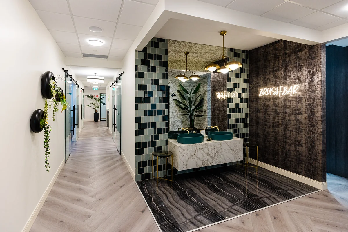
TOOTHbar combines modern digital workflows with a family-owned, comfort-focused dental experience in southwest Calgary.
Frequently Asked Questions About Digital Dental Scans
What is a digital dental scan?
A digital dental scan uses a handheld intraoral scanner to capture visible surfaces inside the mouth and create a three-dimensional digital model of the teeth and bite. The model can be reviewed on screen and used in compatible orthodontic and restorative workflows.
Does a digital dental scan use radiation?
The optical surface-scanning function of an intraoral scanner does not use dental X-ray radiation to create the 3D surface model. Dental X-rays and CBCT scans are different technologies used when the dentist needs information that a surface scan cannot provide.
Does a digital scan replace dental X-rays?
No. An intraoral scan records visible surface anatomy, while dental radiographs can provide information about structures that are not visible at the surface. The dentist selects each type of record according to the clinical question being investigated.
Is a digital scan more comfortable than a putty impression?
Many patients prefer digital scanning because it avoids a tray full of impression material setting in the mouth. Research comparing the techniques has generally found digital impressions to be preferred and associated with less discomfort, nausea and unpleasant taste.
How long does a digital dental scan take?
Scan time depends on the area being recorded, the scanner, your mouth and the purpose of the scan. Digital scanning is usually completed during the appointment, and the clinician can review the model on screen immediately.
What can a digital dental scan be used for?
Digital scans can support many orthodontic and restorative workflows, including clear-aligner records, crowns, selected bridges and other custom dental work. They can also provide a visual record that helps patients understand their teeth and treatment plan.
Are digital impressions always more accurate than traditional impressions?
No single impression method is automatically superior in every clinical situation. Modern intraoral scanners can provide accurate records for many common dental applications, but accuracy depends on the scanner, treatment, anatomy and technique. Conventional impressions still have a role in selected cases.
Does TOOTHbar use digital dental scans?
Yes. TOOTHbar's restorative treatment process includes digital scanning where appropriate, and digital records can also support orthodontic, cosmetic and implant-related treatment planning.
Curious what your teeth look like in 3D?
Digital scanning is one of those pieces of dental technology that is easy to understand once you see it in action. Instead of staring at an impression tray, you can look at a detailed model of your own teeth while the team explains what they are seeing and what your options may be.
TOOTHbar is a family-owned dental studio led by Dr. Murray Knebel in southwest Calgary. Book your appointment online, start with a free video consultation if you would rather ask a few questions first, or call the studio at 403-246-1002.